
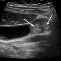
Yup, I’m talking about Adenomyomatosis of the gallbladder.
How much do you really know about this disease? Improve your knowledge and scanning skills by reading more.
Adenomyomatosis is frequently associated with other biliary and pancreatic diseases, such as cholelithiasis, chronic cholecystitis, choledocholithiasis and pancreatitis. Because of this, it is believed that chronic inflammation of the biliary mucosa contributes to the cause of adenomyomatosis. This may also explain why it is more commonly seen after 50 years of age. Time + Inflammation = adenomyomatosis.
So, why is it important to find Adenomyomatosis?
Adenomyomatosis can also cause significant abdominal pain, RUQ pain and hepatic colic, but it can also be asymptomatic. It occurs in both the presence and absence of gallstones. However, it is important to note that adenomyomatosis is a distinct pathology seperate from cholelithiasis and cholecystitis.
Once identified, symptomatic adenomyomatosis is an indication for cholecystectomy, which results in complete disappearance of symptoms in upwards of 90% of patients. Some forms of adenomyomatosis can also be associated with galllbladder carcinoma or leiomyosarcoma, especially in elderly patients.
Which brings up the important question of…
How to find Adenomyomatosis with ultrasound?
This thickened gallbladder wall is most commonly seen in the fundas, but can also occur in the neck area, as well as mid body in a ring-like shape, or generalized over a large area.
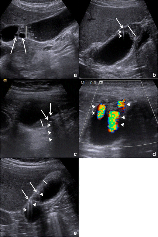
Recommended TIPS for Assessment OF gallbladder FOR adenomyomatosis
2. It is also advised to use a high frequency probe – since the gallbladder fundas can be quite superficial, it is unsatisfactorily evaluated with a 4-5MHz convex transducer. Suspicious findings in this area are better visualized by a higher frequency (7-9 Mhz) linear transducer (link to linear transducer article).
3. Harmonic imaging is also something that will increase the visualization of the gallbladder wall and the detection of Rokitansky-Aschoff sinuses. Use hepatic tissue as a window between the transducer and the gallbladder to also increase image quality.
4. Use multiple windows to assess the anterior wall of the gallbladder very closely for any of the following: thickening (focal or diffuse), areas of irregularity, small cysts and also tiny hyperechoic points (where the bile has crystallized or calcified and creates shadowing), 2D comet tail artifact, color doppler artifact of twinkling, or both.
5. It’s also important to assess any areas of thickened gallbladder wall with color or power doppler, the presence of Rokitansky-Aschoff sinuses will appear avascular, so the presence of any vascularity within a thickened wall should be suspicious for malignancy.
Remember that ultrasound is an operator-dependent imaging modality and the depiction of the gallbladder will vary according to the operators (your) experience.
Take care with gallbladder assessment, remember to always optimize for the organ you are assessing, ask for a second opinion from a fellow tech when you are not sure of something, and use a cine-loop to show any complex or confusing pathology in real-time.
Happy Scanning!
Note:
Images reprinted from the SpringerLink article Gallbladder adenomyomatosis: imaging findings, tricks and pitfalls,(https://link.springer.com/article/10.1007%2Fs13244-017-0544-7) under the terms of the Creative Commons CC BY license.
References and more information:
Gallbladder adenomyomatosis: imaging findings, tricks and pitfalls
https://www.ncbi.nlm.nih.gov/pmc/articles/PMC5359147/#!po=28.7234
https://link.springer.com/article/10.1007%2Fs13244-017-0544-7
Gallbladder adenomyomatosis: Diagnosis and management
https://www.ncbi.nlm.nih.gov/pubmed/28844704
Adenomyomatosis of the gallbladder
https://www.ncbi.nlm.nih.gov/pubmed/1154265
Diffuse Adenomyomatosis Of The Gallbladder: An Infrequent Disease Wth Difficult Preoperative Diagnosis
http://ispub.com/IJS/10/1/6939#
Comments
One response to “Adenomyomatosis: That Other Cause of RUQ Pain”
Interesting blog. Thankyou for sharing this blog.